Health
Entorhinal Cortex and Amygdala Shrinkage May Signal Alzheimer’s Years Before Memory Loss
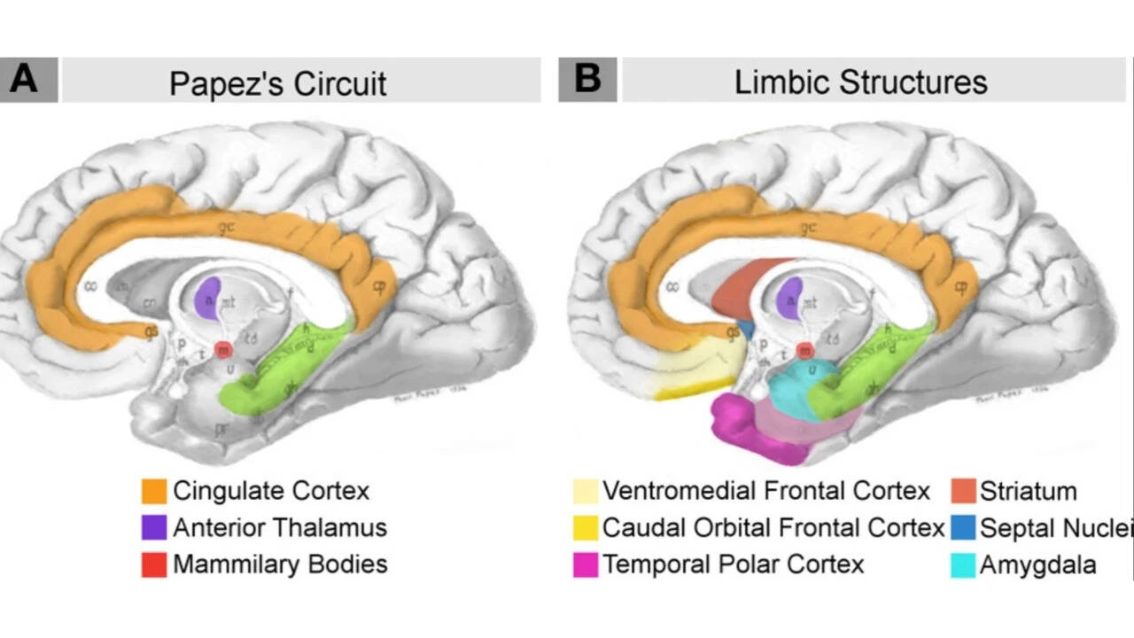
Researchers at Johns Hopkins University, publishing in NeuroImage: Reports in September 2026, found that the entorhinal cortex and amygdala can begin to shrink five to ten years before clinical memory loss appears in Alzheimer’s disease. The study, reported by ScienceDaily on November 14, 2026, examined MRI and PET scans from the Alzheimer’s Disease Neuroimaging Initiative (ADNI) and the Biomarkers of Cognitive Decline Among Normal Individuals (BIOCARD) study, which included participants at various stages of the disease, from asymptomatic to cognitively impaired. Across both cohorts, the entorhinal cortex and amygdala showed greater relative tissue loss during early stages than the hippocampus, a region traditionally linked to memory formation. PET imaging and digital pathology revealed that areas of tissue loss corresponded with abnormal tau protein accumulation, a hallmark of Alzheimer’s pathology. Senior author Michael Miller, a biomedical engineer at Johns Hopkins, noted that these changes could provide a five‑to‑ten‑year window for earlier detection and potential intervention before significant cognitive decline. While the findings represent a promising step toward earlier identification of Alzheimer’s, the researchers emphasize that they are not yet a clinical diagnostic tool and further investigation into molecular changes, particularly tau pathology, is needed.