Health
Rare Genetic Disorder Found in Delhi Man During Infertility Check-Up, Uterus and Undescended Testes Discovered
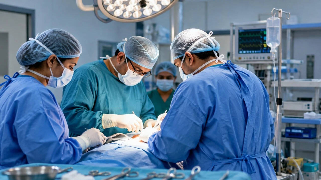
A 26-year-old man who visited RG Hospitals in Rajouri Garden, New Delhi, for treatment of infertility was diagnosed with a rare congenital condition after doctors discovered uterus-like and fallopian-tube-like structures inside his body, according to news agency ANI.
The patient had come in seeking treatment for primary infertility and was diagnosed with azoospermia, a condition marked by the absence of sperm in semen. During examination, doctors also found that both his testes were undescended.
To determine the cause, doctors carried out further evaluations including imaging and genetic testing. An MRI scan revealed a uterus-like structure in the pelvis along with tube-like formations resembling fallopian tubes. Genetic tests showed a 46,XY chromosome pattern, prompting doctors to suspect Persistent Mullerian Duct Syndrome (PMDS) — a rare condition in which Mullerian structures such as the uterus and fallopian tubes persist in individuals with typical male chromosomes and physical traits.
A laparoscopic examination confirmed the presence of a small, underdeveloped uterus and tubular Mullerian structures connected to both testes, which were located inside the abdomen. The testes were found to be severely damaged and atrophied.
Given the patient's age, the long duration of undescended testes, the extent of testicular damage, and an increased risk of testicular cancer, doctors decided to surgically remove both the Mullerian structures and the testes through laparoscopic surgery. The procedure was performed by Dr Susheel Kharbanda, chief urologist at RG Hospitals, Rajouri Garden.
"This was an extremely unusual case because the patient came to us with infertility, and the underlying condition had remained undiagnosed until adulthood. The evaluation showed that he had a persistent Mullerian structure along with both testes located inside the abdomen," said Dr Kharbanda.
He added, "The testes were also severely atrophied, which was an important concern because undescended testes carry a higher risk of testicular cancer. We therefore had to approach the surgery carefully, taking into account both the unusual anatomy and the long-term health risks."
A key finding emerged during microscopic examination of the removed tissue. Both testes showed severe atrophy, and the left testis displayed Germ Cell Neoplasia In Situ (GCNIS), a pre-cancerous change that can progress to testicular cancer. The right testis, while also severely atrophied, showed no signs of GCNIS. Blood tests for common testicular tumour markers returned normal results.
Doctors noted that this case demonstrates how normal tumour-marker levels do not necessarily rule out early or pre-cancerous changes, especially in patients with long-standing undescended testes.
PMDS is typically identified earlier in life, often when a child is being evaluated or treated for an undescended testis. Diagnosis in adulthood, particularly during an infertility evaluation, is rare. In this instance, the investigation into male infertility led to the discovery of both the underlying congenital condition and an unforeseen cancer risk.
The case underscores the importance of investigating unexplained infertility when it occurs alongside bilateral undescended testes. A combination of imaging, genetic testing, laparoscopic examination, and detailed tissue analysis enabled doctors to identify both the condition and the pre-cancerous changes.
RG Hospitals stated that the case was managed through a multidisciplinary approach involving specialists in urology, radiology, pathology, reproductive medicine, and endocrinology. The team relied on imaging, genetic evaluation, minimally invasive surgery, and histopathology to diagnose the condition and manage the associated cancer risk.